
Anatomy for Medical Coder- Circulatory System

Circulatory System for CPC Fresher
Human Circulatory System
Human circulatory system, also called the blood vascular system consists of a muscular chambered heart, a network of closed branching blood vessels and blood, the fluid which is circulated.
The heart is a muscular organ located in the thoracic cavity (mediastinum), between the two lungs and slightly toward the left side. It is approximately the size of a clenched fist and is enclosed within a double-layered sac called the pericardium, which contains pericardial fluid to reduce friction during heart contractions.
The human heart has four chambers:
Right Atrium (RA)
Left Atrium (LA)
Right Ventricle (RV)
Left Ventricle (LV)

Septum
The right and left atria are separated by the interatrial septum,
The right and left ventricles are separated by the interventricular septum.
The atria are separated from the ventricles by the atrioventricular septum.
Valve in Heart
Blood flows through the heart in one direction because of four valves:
Tricuspid Valve: Between the Right Atrium and Right Ventricle
Mitral (Bicuspid) Valve: Between the Left Atrium and Left Ventricle
Pulmonary Valve: Between the Right Ventricle and Pulmonary Artery
Aortic Valve: Between the Left Ventricle and Aorta
These valves prevent backflow of blood.
The heart wall is made of cardiac muscle (myocardium). The ventricles have thicker walls than the atria, and the left ventricle has the thickest wall because it pumps blood throughout the body.
Disease of heart Valve
1. Heart Valve Stenosis
Heart valve stenosis is the narrowing of a heart valve, causing it to not open completely. This reduces blood flow through the valve and makes the heart work harder to pump blood.
Example: Aortic stenosis, Mitral stenosis.
2. Heart Valve Regurgitation (Insufficiency)
Heart valve regurgitation is the leakage of blood backward because the heart valve does not close completely.
Example: Mitral regurgitation, Aortic regurgitation.
3. Heart Valve Prolapse
Heart valve prolapse is a condition in which the valve leaflets bulge (prolapse) backward into the upper chamber (atrium) during heart contraction. It may prevent the valve from closing properly and can lead to regurgitation.
Heart Conduction System
The heart conduction system is the heart's electrical system that generates and conducts electrical impulses, causing the heart muscle to contract and pump blood.
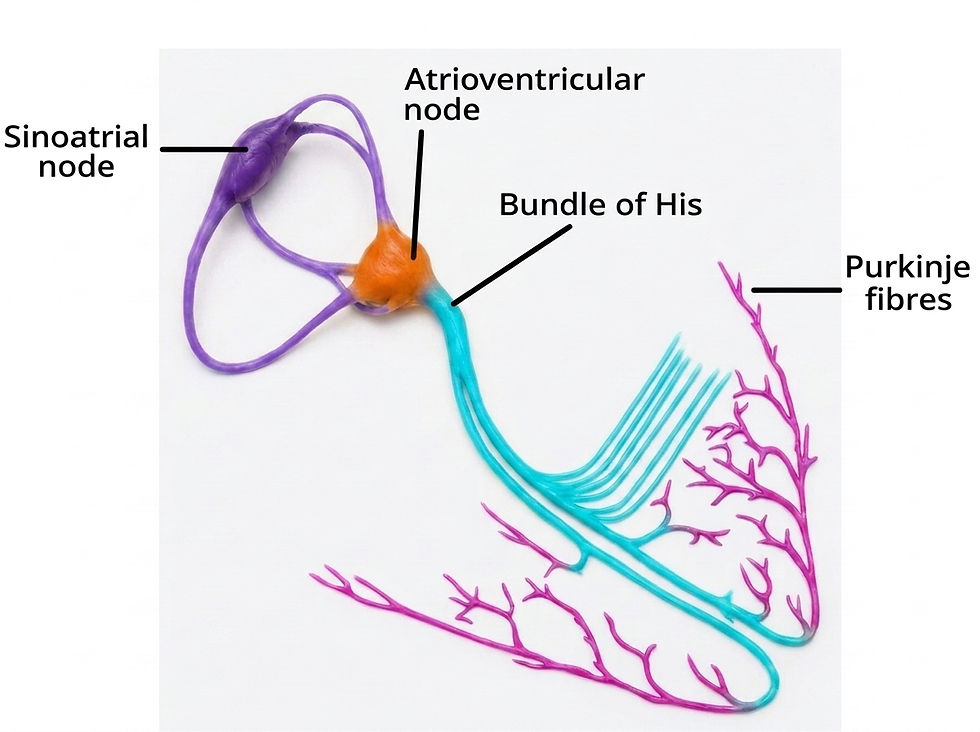
Components of the Heart Conduction System
1. Sinoatrial (SA) Node
Located in the right atrium.Known as the natural pacemaker of the heart.Generates electrical impulses.
Normal rate:
60–100 beats per minute
2. Atrioventricular (AV) Node
Located between the atria and ventricles.Receives impulses from the SA node.Delays the signal briefly so the ventricles can fill with blood.
3. Bundle of His Carries the electrical signal from the AV node into the ventricles.
4. Right and Left Bundle Branches
Run along the interventricular septum.Conduct impulses to the right and left ventricles.
5. Purkinje Fibers
Spread throughout the ventricular muscle.Cause the ventricles to contract.
Disease of Cardiac conduction system
1.Atrial Fibrillation (AFib) -Abnormal electrical activity in the atria.
2.Heart Block -Electrical signals are delayed or blocked.
Examples:
First-degree AV blockSecond-degree AV blockThird-degree (complete) heart block
3.Sick Sinus Syndrome - SA node does not function properly.
4.Ventricular Tachycardia (VT) -Rapid rhythm arising from the ventricles.


Comments